Updated March 28, 2022
New treatments for mast cell tumours have given dogs better chances than ever before. Here you can find out how to recognise a mast cell tumour and what to do if your dog has one.
Just a warning: this is no one-size fits all treatment!
What Is A Mast Cell Tumour?

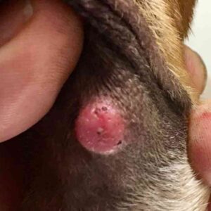
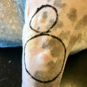
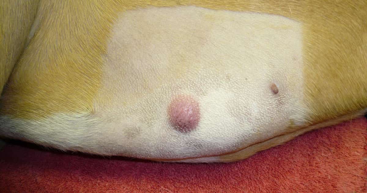
Mast cell tumours (MCTs) are the second most common malignant cancer of dogs, and the most common on the skin. They can vary from a cyst-like lump under the skin to a nasty red raised mass, and affect up to 20% of dogs.
Certain breeds are more likely to develop mast cell tumours, but any dog can get them. The most at risk are:
- Boston Terriers
- Boxers
- Golden Retrievers
- Labrador Retrievers
- Pugs
- Staffies
Due to where mast cells are generally found, most tumours in dogs are located either on or just under the skin. More rarely they can be internal in the liver or spleen.
The pictures here and above give a good idea of what MCTs look like. However, there’s no way to be certain without a checkup and testing.
Diagnosis Of Mast Cell Tumours

MCTs are one of the rare tumours where it’s possible to make a rapid diagnosis at the first visit. A simple fine needle aspirate (FNA) or needle biopsy is all it takes. Once stained and examined by your vet or a pathologist, it can show the characteristic granules inside the cells.
A better, but more invasive test is the incisional biopsy. This is where a small piece is cut out for laboratory analysis. Unlike an FNA, this will require at least a sedation and local anaesthetic. I’ll comment later on when this might be a good idea.
When vets are concerned that the tumour could have spread, they will recommend an ultrasound of the abdomen. They will also want to FNA and possibly remove local lymph nodes.
The benefit of all these tests is that we end up knowing a lot more about the tumour before we try to treat it.
Treatment Of Mast Cell Tumours
There are now four ways to treat mast cell tumours in dogs:
- Surgical removal
- Chemotherapy & radiotherapy
- Tyrosine kinase inhibitors
- Intratumoural injection
The choice of treatment will depend on the tumour’s location, its grade and the owner’s wishes. I’ll go through each in turn.
Surgical Removal
Whenever possible, surgical removal remains the treatment of choice. For solitary MCTs, removal is usually curative. This is possible for the vast majority of dogs. The local lymph node is also removed at the same time if there is suspicion of spread.
However, there are still two related decisions: whether to biopsy and how big the margins need to be. Margins are the rim of normal tissue we remove in order to take out microscopic tumours away from the main mass.
The size of the margins depends on the grade, or aggressiveness of the tumour. Therefore, a biopsy first is always a good idea, and especially in places where a larger margin is going to require planning or referral, such as for high-grade tumours on the legs or head.
On the other hand, for owners wishing to save the extra step, a biopsy is less important in areas where taking a good margin will be easy, like the first picture. This is especially true if the mass has been growing slowly and not causing any trouble.
Margin Size
There is a lot of debate about how big the margins need to be. The current consensus is called modified proportional margins where the margin is the same as the diameter of the tumour, with a minimum of 5mm, a maximum of 2cm, and one fascial plane deep.
Regardless of how successful we think the surgery was, the surgical site and local lymph nodes should always be watched afterwards. MCTs are well known to regrow even after supposedly ‘complete’ removal.
Lab Analysis
The main thing is to always get the tumour graded and the margins checked by a pathologist. This costs around $200 but is money well spent. Then if tumour is found near to the edges, or the MCT is high grade, further steps can be taken.
The decision to re-operate is a tricky one. Even with contaminated margins, low grade MCTs tend not to recur, and intermediate grade tumours only regrow around 33% of the time. I would make this decision based on how practical a second surgery will be.
High grade tumours have a strong tendency to recur, and survival is greatly enhanced by a post-operative course of vinblastine. This can also be a good idea for lower grade MCTs with incomplete removal or lymph node involvement.
Non-Surgical Options
There are at least three reasons why surgery can be the wrong treatment:
- A high grade tumour is in an area where removal isn’t possible, such as the head, legs or around the genitalia
- The dog cannot be safely put through the anaesthetic or surgery
- The tumour has spread elsewhere
These are the dogs who need the next three options.
Chemotherapy & Radiotherapy
Chemo is helpful for certain dogs, but results can be disappointing. A combination of vinblastine and prednisolone +/- lomustine achieves some form of improvement in around 50% of cases. Even if suppressed, tumours will tend to regrow in time.
The use of radiation on the tumour has given improved responses, but it’s not available everywhere. Removal of the primary tumour before chemo also has some benefit even in cases where the disease has spread.
Chemo is probably most useful for tumours that have spread too widely to be able to control the individual masses any more. This is also true for the next treatment.
Tyrosine Kinase Inhibitors
The first major breakthrough in MCT treatment was the development of tyrosine kinase inhibitors (TKIs). The two drugs registered for dogs are toceranib (Palladia®) and masitinib (Masivet® or Kinavet®, not available in Australia).
TKIs are much easier to administer, and can be given at home. Sadly, however, results for these drugs are not much better than chemotherapy, and often at significantly greater cost. Side effects are also generally more of a problem.
All this being said, there are many dogs for whom a TKI has allowed a significant extension in lifespan. The problem we currently have is not knowing how to predict the response before starting.
Intratumoural Injection & Stelfonta
The latest development is the use of injections directly into the MCT. This is only suitable for dogs with small numbers of lumps that are easy to inject. There have been many drugs tried, but only two seem to work.
Triamcinolone is a potent cortisone drug, and may work to suppress or shrink MCTs. There is minimal evidence, but it’s inexpensive and worth a try if funds do not allow for anything else.
Tigilanol tiglate (Stelfonta®) is a new drug derived from a Queensland rainforest tree, which is ironic considering we seemed to be the last to get it. When injected into a MCT, it caused complete remission by 28 days in 73% of cases. Around half of the remaining 27% achieved remission with a second dose and only 7.5% of all the responders suffered a relapse.
Stelfonta is registered in many countries for:
- the treatment of non-metastatic MCT anywhere on the skin surface
- the treatment of non-metastatic MCT under the skin at or below the elbow or hock (in trials, injections higher up caused severe complications)
This only applies to MCTs that cannot be surgically removed and have not spread.
This is a great addition. While surgical removal is still best when possible, Stelfonta can treat MCTs that up to now have required amputations or extremely disfiguring surgery. If you’re interested in Stelfonta, please also scroll down to read about its side effects.
And that’s it. I’m sorry for such a complicated story, but this is the reality of treating mast cell tumours properly. And there will still be some cases that don’t suit any of these treatments. For these, don’t panic; I have a patient right now who’s one year out from a diagnosis of untreatable MCT. He’s doing well and this could be your dog too.
However, one thing always remains the same: the earlier we see tumours, the better the chances. A small lump anywhere can usually be removed, but once they grow you’ll quickly run out of easy options.
We’re lucky to be living in an age of innovations, and so are your dogs. But we still hope you won’t need them.
Also Read: Pictures of Common Lumps Found On Dogs Skin | Types Of Lumps Found Under The Skin
Image at the start by Joel Mills, CC BY-SA 3.0, via Wikimedia Commons.
Have something to add? Comments (if open) will appear within 24 hours.
By Andrew Spanner BVSc(Hons) MVetStud, a vet in Adelaide, Australia. Meet his team here.
References
- Case, A., & Burgess, K. (2018). Safety and efficacy of intralesional triamcinolone administration for treatment of mast cell tumors in dogs: 23 cases (2005–2011). Journal of the American Veterinary Medical Association, 252(1), 84-91
- Chu, M. L., Hayes, G. M., Henry, J. G., & Oblak, M. L. (2020). Comparison of lateral surgical margins of up to two centimeters with margins of three centimeters for achieving tumor-free histologic margins following excision of grade I or II cutaneous mast cell tumors in dogs. Journal of the American Veterinary Medical Association, 256(5), 567-572
- Grant, J., North, S., & Lanore, D. (2016). Clinical response of masitinib mesylate in the treatment of canine macroscopic mast cell tumours. Journal of Small Animal Practice, 57(6), 283-290
- Hahn, K. A., Legendre, A. M., Shaw, N. G., Phillips, B., Ogilvie, G. K., Prescott, D. M., … & Hermine, O. (2010). Evaluation of 12-and 24-month survival rates after treatment with masitinib in dogs with nonresectable mast cell tumors. American journal of veterinary research, 71(11), 1354-1361
- Jones, P. D., Campbell, J. E., Brown, G., Johannes, C. M., & Reddell, P. (2021). Recurrence‐free interval 12 months after local treatment of mast cell tumors in dogs using intratumoral injection of tigilanol tiglate. Journal of veterinary internal medicine, 35(1), 451-455
- London, C. A., Malpas, P. B., Wood-Follis, S. L., Boucher, J. F., Rusk, A. W., Rosenberg, M. P., … & Michels, G. M. (2009). Multi-center, placebo-controlled, double-blind, randomized study of oral toceranib phosphate (SU11654), a receptor tyrosine kinase inhibitor, for the treatment of dogs with recurrent (either local or distant) mast cell tumor following surgical excision. Clinical Cancer Research, 15(11)
- Selmic, L. E., & Ruple, A. (2020). A systematic review of surgical margins utilized for removal of cutaneous mast cell tumors in dogs. BMC veterinary research, 16(1), 1-6
- Smrkovski, O. A., Essick, L., Rohrbach, B. W., & Legendre, A. M. (2015). Masitinib mesylate for metastatic and non‐resectable canine cutaneous mast cell tumours. Veterinary and comparative oncology, 13(3), 314-321
- Weishaar, K. M., Ehrhart, E. J., Avery, A. C., Charles, J. B., Elmslie, R. E., Vail, D. M., … & Thamm, D. H. (2018). c‐Kit mutation and localization status as response predictors in mast cell tumors in dogs treated with prednisone and toceranib or vinblastine. Journal of veterinary internal medicine, 32(1), 394-405
Side Effects Of Stelfonta
The major side effect of Stelfonta is the development of an open wound at least the size of the tumour, which can also be painful for a few days. In rare causes, these wounds are very big indeed. You can see pictures of some in the USA product insert.
The wounds generally heal without complications and leave minimal scarring or disfigurement. Wounds appear to be larger when the tumour has spread, so checking the local lymph node before treatment seems almost essential.
Another potential adverse effect is caused by the release of histamine from mast cells. Deaths occurred in early trials, but these seem to be prevented by pre-treatment with steroids, antihistamines and antacids. Therefore this is also essential.
| Adverse Reaction | STELFONTA 1st Treatment (n = 117) |
| Wound formation | 110 (94.0%) |
| Injection site pain | 61 (52.1%) |
| Lameness in treated limb | 29 (24.8%) |
| Vomiting | 24 (20.5%) |
| Diarrhoea | 24 (20.5%) |
| Low blood protein | 21 (18.0%) |
| Injection site bruising/ erythema/edema/ irritation | 20 (17.1%) |
| Not eating | 14 (12.0%) |
| Regional lymph node swelling/enlargement | 13 (11.1%) |
| Rapid heart rate | 12 (10.3%) |
| Weight loss | 12 (10.3%) |
| Cystitis | 10 (8.6%) |
| Dermatitis | 9 (7.7%) |
| Personality/behavior change | 8 (6.8%) |
| Infection at injection site | 8 (6.8%) |
| Fast breathing | 7 (6.0%) |
| Pruritus | 6 (5.1%) |
| Lethargy/Depression | 6 (5.1%) |
| Pyrexia | 3 (2.6%) |


Great article Andrew. I have a spoodle, who’s just had a high grade mast tumour removed from the side of his rear toes, whole toe was removed. Mast cells found in the closest lymph node a week later. Is it worth removing the lymph node and treating original toes area with Stelfonta? Would love to know your thought.
Hi Anita. Without pretending to be an expert, I think it’s fairly clear that the local lymph node should be removed but there is no benefit in using Stelfonta if the mass itself has been removed. This may be a good case for referral to an oncologist.
Thanks Andrew, we have a follow up early next week for next steps. I appreciate your insights.
Great, thank you so much for this. My little terrier cross dog has had a recurrence of her MCT on her hind leg (after having previous surgical removal x 2). Given her heart condition Stelfonta looks to be her best option. Get you please advise the likely cost of this treatment? I need to decide if I’ll need to trade a kidney for it. Thank you so much!!
Hi Nat. I’m not sure about the price of the drug – I can remember when we looked in the past though that it is surprisingly expensive. Your vet should be happy to get you an up-to-date price.
Thanks Andrew. I’ve since been quoted >$900 for the injection, $1300 for whole procedure (hospitalisation, sedation, etc) but if it gets my Lucy cancer free then it’s worth it 🙂
Thanks Andrew. A very comprehensive article.
Hi my 10yr old ridgeback has a 3” dia flattish lump below the skin at the side of his chest just beside his right leg, armpit area. Will a calcium chloride injection to the lump reduce it as surgery at his age is a risk?
Regards
Anil
Hi Anil. Calcium chloride injections are a discredited form of treatment and no doubt extremely painful. Definitely not recommended.
My 7 month old kitten had a pinnectomy because of high grade mast cell tumor (mitotic count of 8, no splenic or lymph node involvement as the oncologist did needle aspirations and pathology to ensure), fast forward 11 months and he has another on his chin (currently waiting biopsy results). Have you heard of any trials in felines for stelfonta?
Hi Siobhan. No I haven’t, but it also wouldn’t be appropriate for a case like this on the head.
Oh ok, I thought it was ok for cutaneous on head just not sub cutaneous
It’s more that this is a case of multiple masses in a tricky location in an off-label species. There is no easy solution for a case like this so if your vets consider it worthwhile, you should certainly listen to them.
How often do you see a cat with Mast Cell Tumours?
You removed one from my cat, Banjo, a few years ago.
Thanks to your prompt action he is doing well for his age, with no recurrence of MCT.
Hi Deidre – Banjo is the only cat we have ever seen one on! They are very rare and I don’t expect to see another one in my career. Trust Banjo!
My 10 month old GSD/Lab mix was diagnosed with an MCT on/in his nostril. The vet treated the tumor with Stelfonta but the pup required a 2nd treatment after a 30 day exam today. The vet now wants to put my pup on a Vinblastine chemo regimen of 8 doses (weekly for 4 weeks then the remaining 4 doses every 2 weeks). A scan of his abdomen and aspiration of a swollen lymph node was clear. Is chemo the recommended protocol after 2 rounds of non-metastatic (hopefully) MCT?
Hi John. Probably the only other reasonable alternative is surgical removal, which would be disfiguring. However this would also probably be the only truly curative option. You could certainly get a second opinion with a surgical specialist and see what they think of the outcome.
My GSP/Black Lab mix has just been diagnosed with a MCT on his lower front leg. So far it has not spread elsewhere and if everything else checks out he will be getting the Stelafonta injection ASAP.
Good Luck to you and yours.
Interesting to see the comment on one of the side effects of Stelfonta being the release of histamines that can be prevented by pre-treatment with antihistamines and antacids. This is exactly the treatment outlined in the Tagamet/Benadryl Cancer Remission Protocol for dogs ( see Facebook page of same name) which has had some extraordinary results in treating MCTs and other cancers in dogs.
Hi Pam. Thanks for reading to the end! Most mast cell tumours are not actively releasing histamine, it’s more that it becomes a risk when you disturb them. As for the protocol you describe, it may help, but it’s certainly not evidence-based, and like most chemotherapeutic options, it may only work for a limited time before relapse occurs if at all. One of the big problems with assessing efficacy of treatments of MCTs is that their behaviour even when untreated is unpredictable.
My dog has one on his ankle.he seems ok loves to take longer walks..size of marble red raw looking and blk crusty on other side
Hi Katrina. Mast cell tumours are not painful, so he will seem okay for a while yet. The trick is to cut it out before it gets too big.
Great article, thank you.